David Pigott MD, RDMS, FACEP, John Gullett MD, Chris Johnson MD PGY-2
UAB Department of Emergency Medicine
Case Presentation
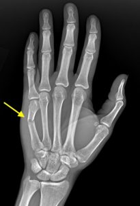
28M presents 3 days after punching a table with left hand pain. Radiographs shows an angulated mid-shaft 5th metacarpal fracture.
To facilitate closed reduction at the bedside, an ultrasound-guided ulnar nerve block was performed in the left forearm.

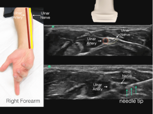
US landmarks for the ulnar nerve block include the ulnar artery and adjacent ulnar nerve. In this clip, the needle is seen depositing lidocaine just superficial to the ulnar nerve. An anechoic “halo” of anesthetic can be seen surrounding the nerve. Note the ulnar artery just to the left and deep to the highly echogenic nerve in the clip.
Although the nerve block was successful, the fracture was unstable and decision was made by consulting Plastic Surgery to take the patient for ORIF the following day. The patient underwent ORIF of his left 5th metacarpal as planned with a plate and screws, and was discharged in good condition.
References
Arun Nagdev, Director of Emergency Ultrasound at Highland Hospital in Oakland, CA, and nerve block guru, gives a great how-to guide for ulnar nerve blocks on ALiEM.
https://www.aliem.com/2016/01/trick-of-the-trade-patient-positioning-for-ultrasound-guided-ulnar-nerve-block/