David Pigott MD RDMS FACEP, Chris Greene MD MPH, Andi Wright MSN FNP-BC
UAB Department of Emergency Medicine
30M with no significant past medical history presents with right posterior ankle pain after playing basketball. He states he came down on his right foot and felt a pop in his right ankle and felt like someone hit him with a baseball bat. He has not been able to bear weight since the injury. On exam, the patient was very tender over the superior aspect of the posterior heel and was unable to plantar flex the right foot.
Point-of-care ultrasound (POCUS) was performed using the linear-array transducer:
The Achilles tendon was visualized in the posterior ankle as well as its insertion site onto the calcaneus. The Achilles tendon was noted to have a slack appearance with obvious disruption of the tendon with an associated echogenic hematoma. Note the location for transducer placement in the diagram.
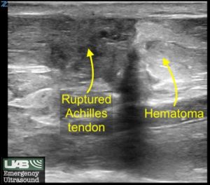

The patient was diagnosed with an Achilles tendon rupture, placed into a posterior splint in equinus configuration (approximately 30° plantar angulation) to offload the Achilles tendon and avoid further traction on the severed tendon.
When tendon ultrasound is performed, it’s important to recognize the appearance of ultrasound artifacts specific to tendon imaging. Anisotropy is an angle-generated artifact. It is produced in tissue that contains multiple, parallel linear sound interfaces (e.g., tendons, ligaments) that lead to the preferential reflection of the beam in one direction.
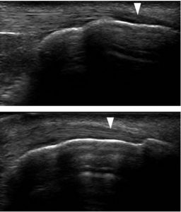
As seen in this example, in the upper image, anisotropy occurs as the fibers of the tendon make an oblique angle to the ultrasound beam near the tendon insertion (arrowhead). This change in the reflection of ultrasound waves leads to a darker appearance of the tendon which may be mistaken for tendon injury. In the lower image, slight change in the ultrasound transducer angle eliminates anisotropy, allowing the entire length of the tendon to be visualized clearly (arrowhead).
A good guide to improving your musculoskeletal ultrasonography is available here:
Arend CF. Top ten pitfalls to avoid when performing musculoskeletal sonography: what you should know before entering the examination room. Eur J Radiol. 2013 Nov;82(11):1933-9.
Copyright permission has been obtained from the UAB Department of Emergency Medicine Division of Ultrasound, Dr. David C. Pigott MD Co-Director of UAB EM Emergency Ultrasound