David Pigott, MD, RDMS, FACEP
Co-Director Emergency Ultrasound, UAB
This 31F patient presented to the UAB Emergency Department with 4 hours of worsening lower abdominal pain in the setting of early pregnancy. She was 7 weeks pregnant by dates. Per EMS, she was tachycardic to the 120s and on arrival was hypotensive, with SBP in the 70s, and diaphoretic.
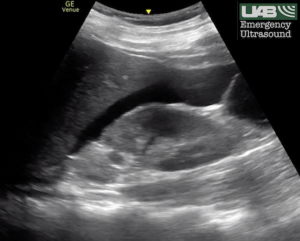
The patient was started on IVF bolus and bedside ultrasound was performed. FAST exam showed a large stripe of free fluid in Morison’s pouch and no clear IUP was seen. (Below)
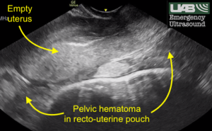
Transvaginal ultrasound was then performed, demonstrating a large pelvic hematoma and an empty uterus. Do not be misled by the relative absence of free fluid in the pelvis. In patients with ruptured ectopic pregnancy, pelvic hematoma rapidly forms, as seen in this case. Anechoic liquid blood may be difficult to detect in this setting. (Below)
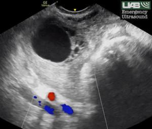
A simple-appearing right adnexal cyst was also seen. Note the follicles within the normal ovary adjacent to the cyst. The internal iliac artery and vein are seen deep to the ovary on this color Doppler scan. This finding does not represent an ectopic pregnancy. It was an incidental finding on Transvaginal ultrasound. (Below)
Gynecology was immediately consulted and resuscitation continued with 2U PRBC and 2U plasma. After Gynecology evaluation, the patient was taken emergently to the OR where a diagnostic laparoscopy was performed. Approximately 1 L hemoperitoneum was noted as well as a ruptured left ectopic pregnancy. Left salpingectomy was performed and hemoperitoneum was evacuated. The patient was discharged in good condition the next morning. She has since returned for post-operative nausea and vomiting but her course has otherwise been benign.
Dr. DeLaney, UAB Associate Professor of Emergency Medicine, and Dr. Brian Bauerband, former UAB EM Chief Resident, published a great summary of potential pitfalls of first trimester sonography on ALiEM.
ALiEM also has a brief guide to TV sonography available here.
For more in-depth information on ED sonography in the evaluation of first trimester pregnancy:
Panebianco NL, Shofer F, Fields JM, Anderson K, Mangili A, Matsuura AC, Dean AJ. The utility of transvaginal ultrasound in the ED evaluation of complications of first trimester pregnancy. Am J Emerg Med. 2015 Jun;33(6):743-8.
Stein JC, Wang R, Adler N, Boscardin J, Jacoby VL, Won G, Goldstein R, Kohn MA. Emergency physician ultrasonography for evaluating patients at risk for ectopic pregnancy: a meta-analysis. Ann Emerg Med. 2010 Dec;56(6):674-83.
© permission has been obtained from The UAB Department of Emergency Medicine for use of these images and clinical case report.